A. Introduction
14.11.25
Many thanks to colleague Dr. Paul Gelinsky for the valuable critical review!
The term “artifact” is used in many scientific fields.
The artifact contrasts with unaltered nature. It is:
- – something inauthentic,
- – something created by artificial circumstances or by human hands
- – something faulty.
The philosopher Gorgias of Leontini (Sicily) essentially claimed in one of his theses:
If something existed, we could not recognize it,
at least not distinguish what is real and what is fake.
The definition of artifacts varies greatly across different disciplines.
In archaeology, an artifact is anything that could have been made by human hands (i.e., the interesting part).
In medicine, an artifact is something that incorrectly represents physiological or pathological anatomy;
in other words, something undesirable.
We are not as pessimistic as Gorgias,
but we are still impressed by how many artifacts exist and how difficult it is to separate them from facts.
In the medical field of radiology, the boundaries are set very narrowly.
If one were to define artifacts too broadly, an X-ray image itself could be considered an artifact—
something that does not exist in nature.
This would render the discussion of “fact or artifact” absurd: everything would be an “artifact.”
Thus, in radiology, the definition of an artifact is limited to everything,
that deviates
- – from standardized examination,
- – from anatomy, and
- – from pathology.
There is even a tendency toward an even narrower definition, restricting artifacts to the medical-technical field,
particularly the film laboratory and the specific circumstances of imaging.
Some experts argue that artifacts should only be considered if they occur outside the patient’s body,
meaning only disruptive phenomena external to the patient should be classified as artifacts.
Wikipedia addresses an important aspect under “Artifact (diagnostics)”:
“…artificially created causal relationships due to errors in data collection, evaluation, documentation, or interpretation.”
Most of our artifacts relate to image formation.
An artifact caused by misinterpretation, so to speak, exists in the examiner’s brain and should occur as rarely as possible.
The author concludes that a clear definition of artifacts is not possible.
However, it is also not necessary.
Even with an awareness of an imprecise definition, artifacts can and should be examined.
The fluid boundaries between artifacts and facts should always be questioned.
What significance do artifacts have for the quality of medicine?
Artifacts interfere with the quality of studies.
It is undisputed that many artifacts can lead to misdiagnoses.
One must know and recognize artifacts in order to react correctly and in time (intervene?).
This preserves quality and saves money.
B. Further Definition Problems
- Can physical phenomena also be classified as artifacts when they lead to undesired (apparent) impairments of images? – Geometric magnification factor, – geometric blur, – partial volume effect, – summation effect? (Can the still lacking knowledge of a law of nature be interpreted as an artifact? The artifact in the examiner’s mind.)
- Is the non-standard projection (see positioning technique) also an artifact?
- Can changes induced by the patient be counted under the concept of artifacts? (From the harmless, provoked deterioration of image quality to Munchausen syndrome). Has there not been an inflation of artifacts in the form of tattoos and piercings in recent decades?
- Is the anatomical variant a (so to speak, genetic) artifact?
- To what extent can pathologies also represent an artifact if an “unnatural” intervention is the cause? For example, trauma and its consequences? One could limit oneself to stating that there are pathologies that exhibit a certain closeness to the concept of an artifact.
In any case, what artifacts and pathology have in common is that they both exhibit an extraordinary variety (a healthy bone varies only slightly from individual to individual within the human species, whereas pathologies and artifacts are diverse). This was demonstrated by the author with the ossification of the first rib. (Schmitt WGH, Pracher W, Medrea M, Kirikuta IC: Manubrium sterni and its jointed connection with the clavicle and the first rib depending on age. Röntgenpraxis 42,193-197 (1989))
C. Various Classifications of Artifacts
Even though the grand solution is still pending, possibilities for classification nevertheless arise (methods of classification). Ten of these are listed below. They are initially very general and then become increasingly specific.
1.- Classification of the artifact according to the breadth of the definition:
Artifact in a narrow or broad sense.
2.- Classification of the artifact according to explainability:
Clear genesis vs. unclear cause
3.- Closely related to this is the classification of artifacts according to avoidability.
Easily avoidable: e.g., electrical discharges on films;
Hardly avoidable: partial volume effect in computed tomography.
4.- Distinction between film-specific artifacts versus those that also occur in digital imaging.
5.- A classification based solely on photographic film: Time and/or place of artifact occurrence within the entire development process;
From the storage of films to the storage of X-ray images.
6.- Also originating from the film era is the division:
Artifacts, unilateral or bilateral film emulsion; this classification was proposed by one of the authors in 1995 in “100 Years of X-rays – 100 Riddles” Schnetztor-Verlag and opens up impressive possibilities for clarifying the causes.
7.- Classification according to the physical phenomena responsible for the artifacts: light, X-rays, mechanical or thermal energy.
8.- White and black artifacts; (in radiological terminology, these correspond to shading and brightening).
This simple classification has the advantage that even less experienced individuals, e.g., trainees, can contribute to statistics.
9.- Classification according to the error value of the artifact:
Harmless, hardly confusable artifacts and those that lead to serious and dangerous misinterpretations.
10.- Classification of artifacts according to the severity of human error:
– unintentional
– well-intentioned, or
– artifacts caused by embarrassing ignorance. (This includes wrong-side, name mix-ups, incorrect projection, etc.),
D. Proposal of a Novel Artifact Encoding
The goal is to find typical artifacts with a simple method (“Ctrl F”) (for comparison purposes, etc.).
This contribution is “unpublished”. Anyone who wishes to modify and publish it is free to do so! He or she has my permission. It is expected that I/we are quoted.
We show many archaic artifacts, many of which come from conventional film development. Nevertheless, we should not forget them. We may be confronted repeatedly with them. At the very least, the old problems educate our minds.
Now, onto the idea. Groups shall be formed that are defined by one or two capital letters. An X is placed in front of the capital letters:
In our collection of cases, specific properties can thus be quickly found using “Ctrl F”.
Example: Search for XA; find all cases of “Anatomical Variants”.
A: Anatomical Variant
BE: Coating affected unilaterally (opposite: BB)
BB: Coating affected bilaterally (opposite: BE)
C: “Computerized technique related” = “Non-film” (opposite: F)
D: Dark Artifacts (opposite: H)
EH: Artifact-generating energy = Heat
EL: Artifact-generating energy = Light
EE: Artifact-generating energy = Electrical discharge
EM: Artifact-generating energy = Mechanics
ER: Artifact-generating energy = X-rays
EN: Artifact-generating energy = Radiation from a radioactive substance (Nuclear Medicine)
FD: Film in depot (unexposed material) artificially damaged
FL: Artifact occurrence during film load of the cassette
FB: Artifact occurrence during film exposure in X-ray
FO: Artifact occurrence on film when opening (i.e., opening) the cassette
(All F in contrast to C)
FE: Disruption of film development
FF: Disruption of film fixation (and washing)
FT: Disruption of film drying
FA: Faulty film storage, archiving
G: Mixed = light + dark artifacts (Opposite of D and H)
H: Light Artifacts (Opposite of D and G)
IB: Iatrogenic artifact despite best intentions
IN: Iatrogenic artifact due to negligence
II: Iatrogenic artifact due to gross ignorance (not M)
J: Projection Error
KJ: Disease value Yes!
KN: Disease value No!
L: Educational Case
MM: Medical-historical malpractice
MR: Magnetic Resonance (NMR = Nuclear Magnetic Resonance Imaging)
N: Natural phenomenon – Physics – Physiology
O: Ordinary = more frequent, well-known artifact (Opposite of R)
P: Pathology (not a true artifact?)
Q: Source Patient: The patient produces the artifact unintentionally ………. (Opposite of T)
R: Rarity (Opposite of O)
S: Hardly avoidable (Opposite of U and V)
T: Deception by patients up to Munchausen syndrome ……. (Opposite of Q)
U: Unavoidable artifact (Opposite of V and S)
V: Avoidable artifact (Opposite of S and U)
W: Broadly defined artifact (Opposite of Z)
Z: (Overly) narrow definition of the artifact (Opposite of W)
If two or more criteria apply, then the X must be repeated accordingly. Each criterion begins with X, so that even in a longer text the criterion YZ can be found, it must be extended to XYZ.
]
1. Capitel. Light artefacts:
Dieser Betrag will die These verteidigen: Es lohnt sich, Artefakte zu zeigen und zu studieren. Genaue Kenntnisse sind notwendig zur Vermeidung. Oftgeht eine Detektiv-Arbeit voraus. Diese sollte früh erfolgen, ehe ein Artefakt in Serie auftritt.
Nebenbei lernt man aus der Fahndung nach Artefakten viel für die tägliche Diagnostik (siehe Beitrag “Pathologie”).
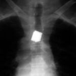
Eine besondere Gruppe sind die verschluckten Fremdkörper, wie sie im folgenden Kapitel ca. ab Abb. 39 gezeigt werden.
Zur Nomenklatur:
Man könnte die “hellen” auch “weiße” Artefakte nennen.
Wir legen uns fest auf den alltäglichen Sprachgebrauch und meiden die klassische röntgenologische Benennung, wo etwas relativ “Weißes im Bild” ein “Schatten” ist.;
Das zweite Kapitel ist den “dunklen” (schwarzen) Artefakten gewidmet, also den röntgenologischen “Aufhellungen”. Diese sprachlichen Residuen aus der Entstehungszeit der Röntgendiagnostik vermeiden wir, da sie zu verwirrend ist. Diese alte Nomenklatur stammt aus der Zeit, als man sich auf die Eindrücke am Durchleuchtungsschirm bezog. Dort ist das Bild im Vergleich zum Film/Computer invertiert.
Wenn wir uns die hellen Artefakte rausgreifen, kann man einige Regeln erkennen:
1.
a) Nur die optimale Belichtung nutzt die Möglichkeiten des Filmes voll aus.
b) In dieser normalen Filmschwärzung sind helle Artefakt (also auch Fremdkörper) wesentlich weniger deutlich, ja diskrete Formen verschwinden scheinbar. Unter der hellen Lampe oder bei elektronischer Aufhellung werden sie aber deutlicher erkannt.
Nur wer wenig Praxis hat, vermutet einen Widerspruch zwischen a und b.
Es ist falsch, absichtlich niedrig zu belichten, in der Absicht Fremdkörper besser zu detektieren.
Die folgenden Aufnahmen wurden regelrecht belichtet aber in diesem Beitrag recht hell abgebildet. Das sind keine fehlerhaften Unterbelichtungen.
2.
Helle Artefakte sind überwiegend Fremdmaterialien, die versehentlich zwischen Focus und Film geraten sind (Außerhalb oder im Patienten).
3.
Die Strahlenschwächung dieser artifiziellen Körper kann absolut und relativ betrachtet werden.
a) Uns Physiker interessiert die absolute Strahlenschwächung.
b) Als praktische Radiologen wollen wir den relativen Wert kennen; wie deutlich kommt das Artefakt heraus, und wie ist der Kontrast?
Zu a) Absolute Strahlenschwächung: Die “Verschattung” eines Objekts ist abhängig vom Material (Strahlenabsorpitonskoeffizient) und Dicke. (siehe WGHS et al.: Was sagen die Houndsfield-Einheiten?)
Zu b) Relative Strahlenschwächung: Der Kontrast kommt zustande durch Unterschied in der Schwächung von Objekt und Umgebung.
Wer in der Radiologie startet soll sich lediglich merken:
Gleiches Objekt wird sehr unterschiedlich dargestellt, je nachdem ob es von Luft oder von Weichteil (Flüssigkeit) umgeben ist.
– Jedes Material dichter als Luft ist “in Luft” schattengebend”, kann also ein “helles Artefakt” erzeugen.
– Das gleiche Material ergibt keinerlei Kontrast in einer Umgebung gleicher Dichte.
– Das gleiche Material wird zum ” schwarzen Artefakt” wenn es von einer Substanz größerer Dichte umgeben ist.
Besser als viele Worte sind einprägsame Beispiele:
1.01 Defekt in der Tischplatte
Hier fehlt das Bild
Kommentar zu 1.01 XBBXHXINXKNXL
Zu Anfang ein Artefakt, der eine Herausforderung darstellt. In gleicher Gestalt tritt er bei ganz verschiedenen Untersuchungen immer wieder auf. Die Kunst besteht darin, ihn im Gedächtnis zu speichern, als Artefakt zu erkennen und nicht in 5 Wochen, sondern am ersten Tag seines Auftretens zu beseitigen. – Wichtiger Fall.
Jeglicher Defekt in der Unterlage hätte die Homogenität gestört und sich möglicherweise abgebildet. Vielleicht wäre dieser Artefakt so gering, dass er lange nicht auffällt.
Hier ist der Defekt auf eine besonders störende Art hervorgehoben:
In einem Defekt der Tischplatte eingetrocknetes BaSO4. Der Kontrastmittel-Fleck wurde auf (fast) jedem Film abgebildet. Es bestand die Gefahr von Serien-Fehldiagnosen.
Wir sind herausgefordert zu erkennen: Diese Struktur habe ich bereits bei einem anderen Patienten und/oder in einer anderen Region in identischer Form gesehen!
Es war ein Bucky-Tisch. Die Kassette befindet sich unter dem Tisch in einer Schublade. Zwischen Kassette und Tisch ist die Bucky-Blende, die Streustrahlen wegfängt. Auf dem Tisch liegt der Patient.
Wenn das Team uneinig ist, hilft nur eine Belichtung ohne Patient, deren Ergebnis normalerweise kein Artefakt zeigen darf.
1.02 Torn silver paper strips from a film pack. While loading the film, it got caught between the film and one of the intensifying screens.
1.03 One of the Classics among Artifacts. Buttons on the Thorax.
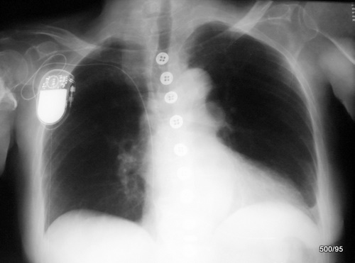
3 XBBXHXKNXO
– Buttons are made of relatively dense material (approx. +1000 HU, not nearly as radiopaque as metal), but they are thin compared to the human thorax; in the upper and middle buttons of the button row, there are distinct artifacts because the thorax is “thinner”, that is, more transparent, in that area.
Their (unfortunately) good contrast also arises from the fact that they are surrounded by air. – A swallowed button (surrounded by gastric juice) would make it more difficult for us.
The lowest buttons are not depicted as well. As we have already mentioned: The mid-field is thicker (denser) in the middle and bottom. One reason is the overlap caused by the heart. This leads to underexposure, which is very dangerous for diagnostics.
I found it unnecessary, dear friends of radiology, to present you with further everyday examples of artifacts. You encounter these daily and know them well:
The finger ring in the X-ray image. Please pay attention next time to the psychology of the X-ray image. Such a ring appears to be “hovering” in front of the image. In reality, the finger is inside the ring; something light (in the sense of white) in the X-ray image always appears to us as being very close to the viewer, whereas something dark appears to be farther away; this suggestion makes correct spatial perception extremely difficult. This suggestion is the source of many misdiagnoses. Even in this case, one would reveal the optical illusion through a second plane.
Another everyday artifact is the braid “pinned up” with hairpins and clips. Naturally, this also includes piercings. Such metal parts should be removed before a proper exposure.
1.04 Misdiagnosis: Infiltrate in the Upper Field
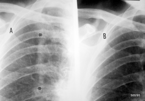
4 XBBXHXINXKNXLXQ
This case is not as simple and not as common as the aforementioned buttons, jewelry, or hairpins.
Overlap of the thorax by a braid (marked in A by 2 black stars).
All materials are radiopaque in surrounding air if they are denser than air. If something is surrounded by air, we must not draw overly extensive conclusions about the nature of the material from the X-ray image. This simple rule has frequently been ignored in the history of radiology. – The phenomenon is also illustrated in the article on Radiation Protection (3.2a).
There was no consensus regarding the benign nature of the change. Therefore, the image was repeated (see image part B) while avoiding the disturbing overlap. The artifact is proven; a pathological lung change in this area is ruled out.
1.05 Striated Densities Projected onto the Cranial Vault: Hairstyle Causes Slight Atypical Superimpositions. Image Underexposed.
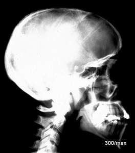
1.06 Boy with Many Curls in the Skull X-ray ?? (Excerpt)
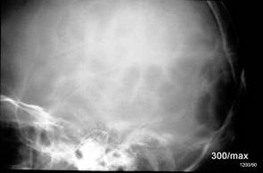
1.05 upper image (try classifying it yourself!) Bicycle accident. No concussion. Frontal hematoma. Otherwise, no clinical findings. The monitoring was carried out carefully. We would have refrained from the X-ray examination.
Diagnosis: Not pronounced superimposition of the skull by braids. An individual hair escapes depiction under these conditions (after all, the human skull is voluminous). Only when “bundled” and surrounded by air do the hairs become visible and create a diagnostic problem.
1.06 lower image XBBXHXKN
The clinical picture is very similar to case 05. The indication is likewise controversial.
The blurred black spots, however, are by no means the curls as laypeople suspected when viewing the X-ray image, but merely a harmless variant in the structure and thickness of the cranial vault. There was no neurological symptomatology, no evidence of increased intracranial pressure, no bone marrow disease.
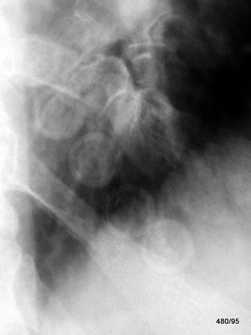
1.07 These calcified patchy opacities are projected onto the neck, mediastinum, thoracic wall, and lung.
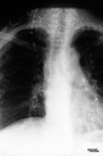
07 XBBXHXKNXLXQ
For calcified lymph nodes, the spots are unusually small, too regular, and too distinct. Even a slight magnification of the X-ray image reveals the ring shape of the small spot opacities; this suggests an artificial origin.
The examination of the patient brought – as so often – clarity. “Multiple braids: colorful beads and rings are woven into the hair.” (Certain pigments and metals could increase the radiation absorption.)
- The braids, unlike in cases 04 and 05, are apparently too thin to provide their own contrast.
- The mentioned beads and rings are large and dense enough to create a marked contrast.
Therefore, the commentator considers the remark “colorful” necessary. For example, the radiation attenuation of a certain plastic can vary depending on the admixture of pigments. (see WGHSchmitt, KH Huebener: Density of wood, plastic, and glass. RöFo.135 (1981) 2512-17)
1.08 Is the Arrow Really Stuck in the Head?
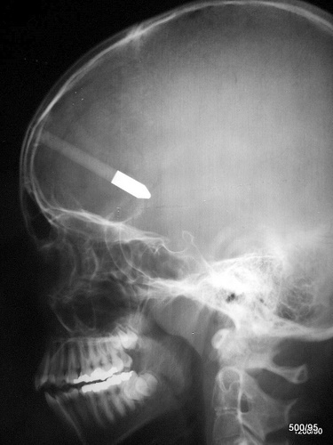
08 XBBXHXKJXLXQXPXW
We knew what was going on. However, we wanted to pose this puzzle: purely theoretically, from a single plane we cannot know. We could assume it with some certainty under a special condition: if the non-metal part of the arrow were isodense to “water,” then it would produce no contrast where it is surrounded by tissue. A very theoretical and difficult argument!
The appearance proved it, and the second plane documented it: the arrow was lodged in the middle of the head, exactly between both hemispheres. The shooter was the patient’s brother. The course after removal of the arrow during surgery was pleasantly uncomplicated and free of any residue, apart from a prolonged period of discord between the brothers.
1.09 Bright or Dark Artifact? Parallel Stripes of the Cranial Vault?
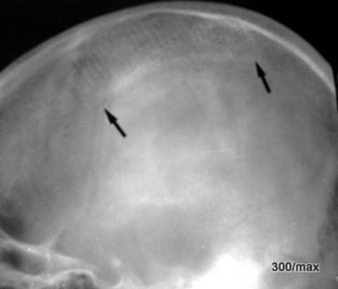
09 Overlap by a comb. (Marked by 2 black arrows). Isn’t it disappointing how our sensory physiology lets us down. Some of us would have sworn that it was a black artifact. Black stripes that somehow appear mysteriously in or on the skull. Once you embrace the idea of a bright artifact and the concept of an overlapping comb, everything becomes quite logical. Even the thin comb can add enough radiation attenuation to the skull to be depicted quite clearly. An unusually harmless artifact, but when it comes to interpretation, even such banalities challenge us.
1.10 Bright or Dark Artifact in the Femoral Neck?
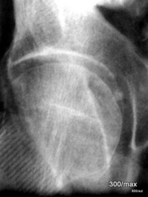
10 XBBXHXINXKNXQ
By now, you have been trained by the previous case and immediately recognize that it is again a comb that overlaps the femoral neck. Again, some are initially convinced that it is a black artifact, but it is an additional structure “with bright tines”.
1.11 The bright grid over the entire breast is an overlap of the cassette cover.
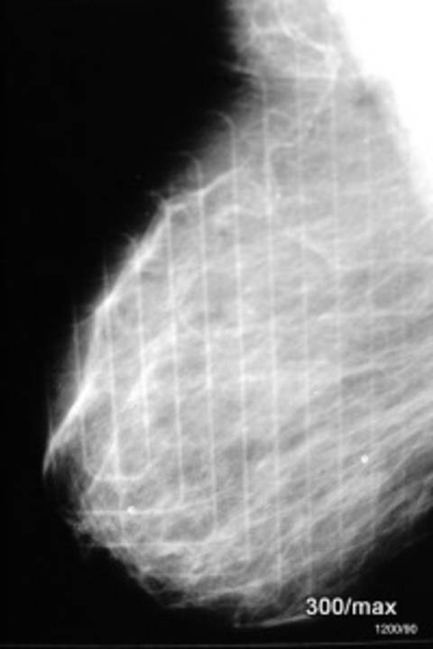
11 XBBXHXINXKN
Front and back of the cassette were swapped during the exposure.
This mammogram is rendered in a very coarse grid; it would not be usable for diagnostic purposes. It was solely about the artifact. In the cassette, the film is in close contact with one or two intensifying screens. One side is completely smooth and faces the patient. The other side, the cassette cover, has a reinforcing profile to improve stability.
Everyone is trained and strives to avoid such an error. Most have never experienced it; I have only once. Some of our examples are from the time when X-ray film was still widely used and chemical processing played a major role. Today, we no longer have to deal with many of these problems. However, we may sometimes be confronted with old techniques. Moreover, it is a good training for the eye and mind.
1.12 Is the Ornamental Form of the Opacities Atypical for a Pathological Process?
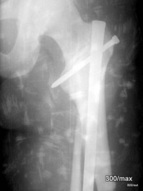
12 XBBXHXINXKNXQ
Yes! The front of the bathrobe was turned to the side and the setting was made, but the back part was not considered.
The silver-shiny thread used contains metal.
Ein ordinary twisted thread is not capable of producing a shadow visible to the eye. However, there is the special case discussed in “Article 05 Pleura I”, demonstrated by E. Pantoja.
We understand the drama of the clinical situation and the reason for the haste. The nail in his proximal femur has dislodged.
1.13 Is that also an article of clothing that overlaps the X-ray image?
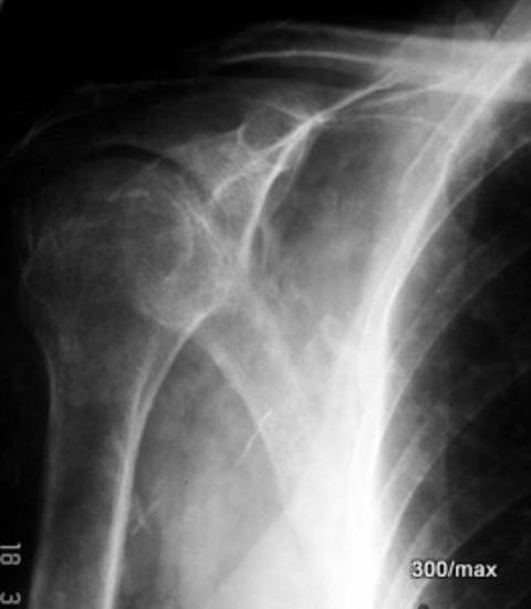
13 XBBXHXINXKNXQ
The assumption that this is an artifact is facilitated by the fact that the bright structures also lie outside the bone. Under the bright lamp, it should be checked whether the shadows extend beyond the soft tissues. If that is the case, the diagnosis “artifact” is particularly straightforward. That is exactly what happened: embroidery.
1.14 Artifact or Cysticercosis?
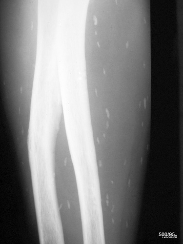
14 XBBXHXKJXLXRXQXPXW
We also initially thought it was an artifact;
the examination of the patient and the entire setup, including the cassette, yielded no results.
What was striking was the restriction of the calcified opacities to the soft tissues. –
It was decided to examine another region of the body; even in the contralateral forearm, identically shaped calcifications were detected in the soft tissue area.
Serologically: Confirmation of the diagnosis cysticercosis.
1.15 Excerpt from a Double Contrast Stomach Examination.
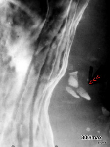
15 XBBXHXINXKNXQ
The extragastric dense opacities (red arrowheads) are artificial; these are barium sulfate spots on a shirt, with the textile structure discernibly indicated.
1.16 Overlap of the Hip Region?
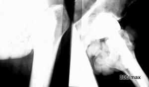
16 XBBXHXINXKN
A large-volume, very dense overlap is present. It is an anus praeter (stoma bag and contents).
The image is severely underexposed. Even with adequate exposure, prior to cleanup there is no chance for a differentiated diagnosis of the hip joint and proximal femur.
1.17 Tamponade of the Left Nasal Cavity (red arrowheads). Indication for the examination unclear.
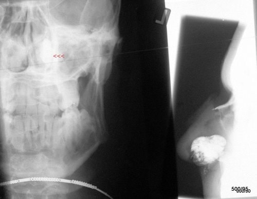
17 XBBXHXIBXKNXL
The textile material alone would not explain the radiation attenuation; but it is soaked with an ointment that significantly reduces the radiation.
Here, the primary concern was to treat the complication of the injury: to stop the nosebleed. The X-ray examination was requested to view more of the bone. One must frankly say that the X-ray is not the most important factor in the question of a “nasal fracture”. Often, the X-ray image is not necessary. The ENT findings are very important: if there is a hematoma in the nasal septum, then a nasal bone fracture likely needs to be realigned (reduced). This is done under anesthesia.
The image here was, so to speak, a by-product—not even a pretty one, but in my opinion, interesting.
1.18 A layered intrathoracic calcification or an artificial overlap?
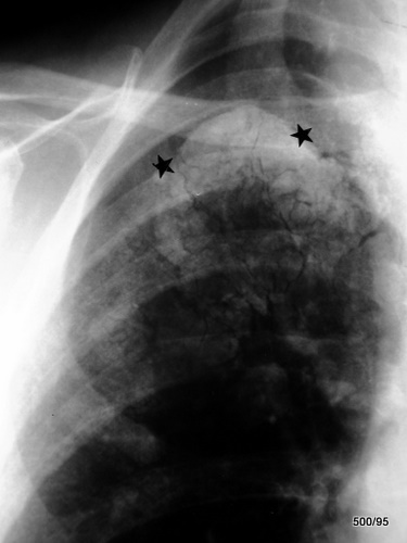
18 XBBXHXIBXKN. It doesn’t quite match an intrathoracic calcification. Pleural calcification can be very pronounced; in a tangential layer it then produces a very distinct bright line. In the overview, the calcification layer is usually not thick enough to be seen. Could it be a pleural cavity filled with air above the calcification? That does not fit with the fact that we see nothing of a parietal pleural sheet. See the 4 articles Pleura 04–07. It is a misconception that the parietal pleura does not calcify. In short, the simplest explanation is often the best: take a close look at the patient. This is an overlapping ointment dressing!
For the explanation of the image it is important to note that the dressing has absorbed a large amount of ointment. This ointment has a radiation-attenuating property (perhaps due to its zinc content). Without the ointment, the dressing material would probably not be discernible.
Thus, it is very likely not a lung process and not a pathological change of the thoracic wall. We hope that we can remove the dressing cleanly and perform a follow-up examination that provides a wealth of information and, among other things, also rules out a pneumothorax at the right apex.
1.19a+b The case dates back 5 decades. Breast cancer; T4 ulcerated. Status post radiation therapy.
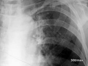
1.19b
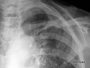
19a XBBXHXKJXPXW
Large scar plate with calcifications of the thoracic wall.
The therapeutic options with photons and electrons, as well as therapy planning, have meanwhile fundamentally changed. – An osteolysis of the clavicle is mentioned. No accident is possible, nor are radiation-induced sequelae.
19b Second image: 7 months after the first; the scabby calcifications are even somewhat more pronounced; blurring of the dorsal upper margin of the 4th rib is highly suspicious for osteolyses (see article 08 “Bone Metastases”).
It is correct that the case does not belong to artifacts in the strict sense. It could be classified in Chapter 4 (Iatrogenic Changes). The scarring and calcifications of the thoracic wall will hopefully remain a thing of the past.
1.20 Thorax with Glasses. In addition to the artifact, there is also a true “pathology” in the area of the right lung and pleura.
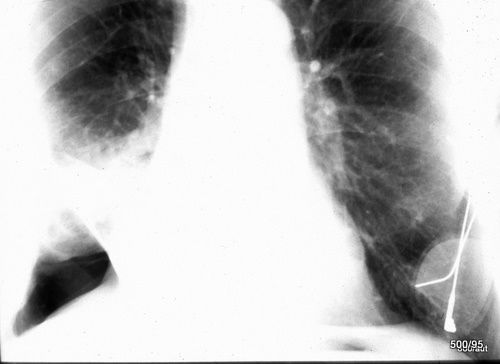
20 XBBXHXINXKNXO. First, regarding the glasses. The relative radiation attenuation of the eyeglass lenses: the good contrast is achieved by the difference in attenuation between the object and its surroundings. “Eyeglass lenses surrounded by air cast a more pronounced ‘shadow’ than when they are surrounded by water. Any material denser than air casts a shadow in air.” Relativity of shadowing. Of course, its absolute radiation attenuation is completely independent of the surrounding medium. The “shadowing” of an object depends on the material and its thickness. Glass is generally somewhat less attenuating than the metal of the temples and slightly more than the plastic of the eyeglass frame; this in turn is slightly more than soft tissue. We could express the attenuation values in HU (see WGHS: What do the Houndsfield units = HU say…). These are trivialities.
In addition, there is an atelectasis in the right lower lung field, apparently a middle lobe atelectasis.
Atelectasis is also frequently mentioned in the article “Pleura and Thorax”. The lung becomes airless and therefore as dense as “flesh”. It is difficult: What is the difference between atelectasis and a pneumonia (lung inflammation)? Both appear as something “bright” (radiological terminology: opacity) on the X‑ray image. Pneumonia is an infectious disease encompassing a large group of conditions. Atelectasis is merely a sign, a symptom of either a bronchial obstruction or a compression of lung tissue. Many factors can be causal.
The structureless area in the right lower field baffles us. Until proven otherwise, it is considered a pneumothorax, possibly following an attempted puncture. I lack the data; this deficiency should be remedied as soon as possible.
1.21 Accident, excerpt from a lateral skull X‑ray. – What is the bright (radiologically: radiopaque) structure in one or both orbital cavities?
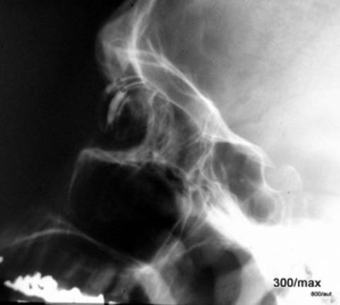
21 XBBXHXL Glass eyes in both orbits. The depicted pneumatic cavities appear well ventilated.
1.22 Same patient, AP; not sufficiently centered and displayed.
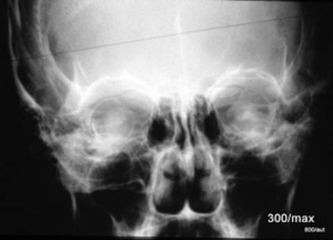
1.22 XBEXHXL
1.23 Another patient. Was an orbital floor fracture suspected?
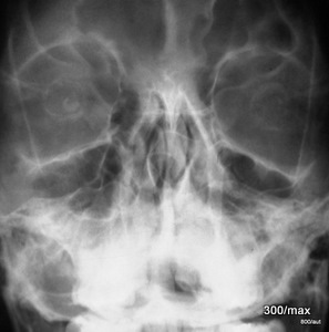
23 XBBXHXL
No evidence of fracture. Jaw sinuses clear.
Also, glass eye prostheses
It seemed important to me to demonstrate various brands and the corresponding diversity of forms.
Incidentally, the image is also suitable to review the linea innominata and, in general, the positioning technique. – If this exposure were intended to depict the jaw sinuses, what was done wrong? Raise the chin further to project the base of the aforementioned sinuses clearly. The linea innominata runs through the lateral quarter of the orbit from top to bottom; it is inconspicuous.
Skull radiography is rightly indicated more critically today.
1.24 An elaborate method to document temperature?
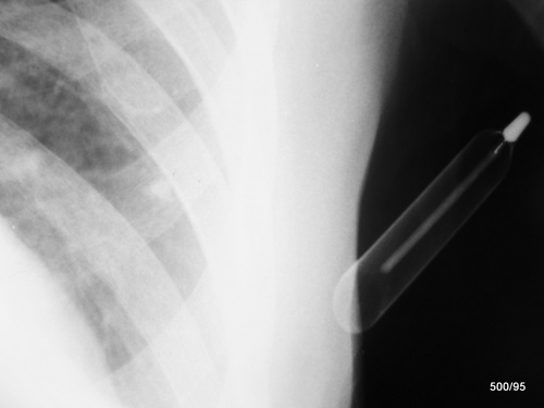
24 XBBXHXINXKNXO
In all the seriousness of everyday life, here is a curiosity. Perhaps an opportunity to ask how the glass tube (e.g., of a thermometer) is depicted on an X‑ray; it is shown most distinctly where it is tangentially hit by the radiation.
When traversed orthogradely, almost nothing of the material is visible, except for the central, wall-thick tube.
This observation applies to all “shells”. There are many examples of these in the body. Therefore, this rule is so important; see the three-part article 04–07.
1.25 Patient after a rollover accident. X‑ray of the elbow. What are the quadrangular bodies?
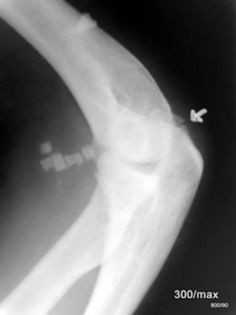
25 XBBXHXINXKNXPXW
Overlap due to splinters from safety glass. These external glass splinters (surrounded by air) produce a much better contrast than those that would be embedded in tissue.
It is therefore annoying that glass splinters are much more difficult to diagnose when it matters. See the next case (although with bottle glass). – Fragmentation at the tip of the olecranon. Age? Clinical details? We know too little here.
1.26 Glass Splinters? Position? (Check Functions!)
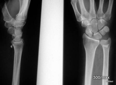
26 XBBXHXKJXLXPXW
Yes! They are located in the soft tissues – distal radius, volar. – Often, the depiction of glass splinters is more difficult. It is easier the larger the glass splinters are, the better they are projected tangentially and the less overlap there is.
We have long moved away from the advice of underexposing such examinations. By brightening with a strong lamp, one can ensure that nothing hides in dark areas of the image; however, in areas that are too bright, information can be irretrievably lost. A seeming exception is the following: when I want to present such images online, I like to make them a bit brighter. Do not be surprised if I frequently present noticeably bright images that would not be accepted by the Medical Authorities.
Overlooked foreign bodies are such an important problem that we will show a similar case again.
1.27 Hand Injury in a Beer Tent. What’s Special?
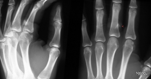
27 XBBXHXKJXLXPXW
Glass splinters ventral to the base of the proximal phalanx of the third finger. The change in position when rotating the hand outward (left image; “beginning supination”) proved the volar location.
Compare the three cases above with a “hypodense” splinter in case 2. 28!
1.28 Granular Material (denser than soft tissues)? What could it be?
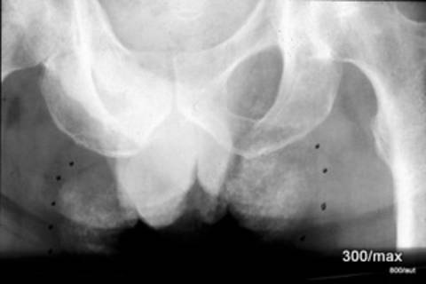
28 It is a “dislocated” sandbag wich should compress a inguinal puncture. .
1.29 Coarse-grained dense material in the area of the thorax and shoulder?
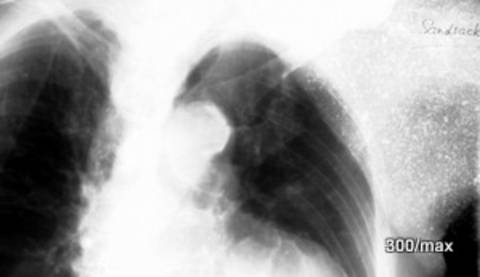
1.29 29 XBBXHXINXKN
Ausschnitt aus einem Thorax a.p. im Liegen. Diagnostik fordert manchmal auch Improvisationen. Hier wurde versucht den verwirrten Patienten mit einem sanften Gewicht zu bewegen, seinen Arm nicht über den Thorax zu legen. Wegen der schwierigen Bedingungen ist auch die Einblendung so ungewöhnlich mangelhaft. Doe Antwort ist also: Sandsack über der Schulter , bis in die Thorax-Region reichend. Trotz Bemühungen unzureichende Diagnostik.
1.30 Polytraumatized Motorcyclist. What happened here?
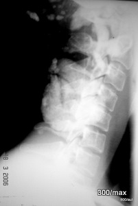
30 XBBXHXINXKNXQ
Patient’s age? 17 years. He tumbled several times and landed in a swamp.
Did that save him from more severe injuries?
In a rush and to obtain orienting information, this exposure was made, but it did no service to anyone.
The original exposure is not underexposed; rather, it was deliberately processed very brightly.
External contamination with sand, mud, and dirt.
Nowadays, in such an accident and at the corresponding clinic, a CT would probably be performed wherever possible.
However, even for that, such artifacts would have to be eliminated. This will cost valuable time, but it will bring diagnostic clarity.
One can understand what the emergency preparedness must accomplish in individual cases, and that these services are rarely appreciated.
In summary: Today, in cases of polytrauma, we favor rapid CT clarification.
The prerequisites are secured vital functions and, in the truest sense, the elimination of such artifacts as shown here.
1.31 Accident of a 15-year-old, avulsion fracture in the area of the spina iliaca inferior?
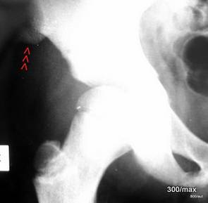
31 XBBXHXINXKNXLXQ
The 15-year-old boy (see growth plate of the greater trochanter) landed on the water surface after jumping from the 3-meter diving board, with the front of his thigh.
Despite this mechanism of injury, there was no clinical finding that would have raised the suspicion of an avulsion of the anterior iliac spine – fact or artifact? –
This exposure is also reproduced with brightening to demonstrate the finding more clearly. – It is remarkable that the patient was not wearing swim trunks, but rather shorts with pockets. – In this “shadow” it was sand in a pocket. –
1.32 Pain in the right midface. Imaging of the paranasal sinuses, rarely used, replaced by sonography.
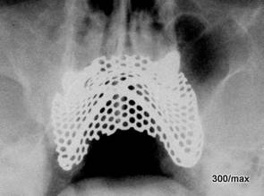
A common (and as often avoidable) artifact.
Right maxillary sinusitis. As a strongly quality-reducing curiosity, a metallic mesh that reinforces a maxillary dental prosthesis.
1.33 Attempt at a thorax AP x‑ray from decades ago
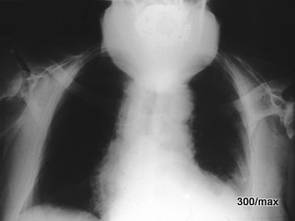
33 XBBXHXINXKNXQ. The unfortunate critically ill patient suffers from a kyphosis that forces the head into an abnormal posture.
The kyphosis results in the peculiar projection of the facial skull and a dental prosthesis; this overlaps with the thorax, which also has completely failed in terms of exposure.
1.34 The key to the diagnosis in this severely underexposed image
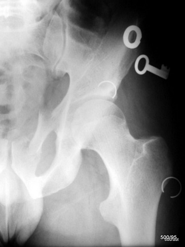
34 XBBXHXIBXKNXQ. One of the real keys is fractured at its “locus minoris resistentiae”; two metal rings are also broken – fragmentation of the superior pubic ramus.
The trauma has therefore not only affected the pelvic bone but even the key ring. The situation of the injured patient may explain the haste during this X‑ray examination. Was the heavy key ring an additional hazard to the victim, or did it absorb energy that provided some protection to the bone? We need not know.
Artifacts usually complicate the diagnosis. Here is a curiosity: the artifact essentially provides an additional clue to the accident mechanism.
1.35 Excerpt from a thorax examination. An old 5-mark coin in the X‑ray. What is on the left side of the image?–
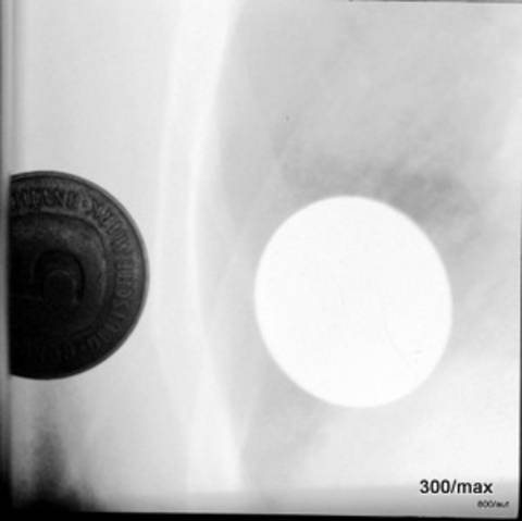
35 XBBXHXKNXQ
The image is, as often in this collection, reproduced very brightly, much brighter than would be permitted in the original. It dates from the year 2000.
The coin in the pocket of a shirt. Hence the X‑ray image of a coin. Of course, a silly mistake. – On the left, another coin was affixed to the film for demonstration purposes.
This is how the coin is depicted both optically and radiologically.
With the large focus-film distance of the thorax examination and such a “plate-near” foreign body, the geometric magnification (not infrequently a problem in precise measurement) is very small. (Compare 38).
1.36 Overlap of the hip region by a magnetic tape cassette.
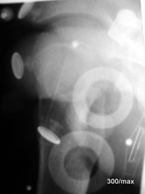
36 XBBXHXKNXNXOXQ
The wound-up tape causes a surprisingly strong radiation attenuation (due to the cumulative iron content). The non-wound portion of the tape is barely discernible.
The plastic housing is also surrounded by air, which should enhance its contrast; however, the attenuation values of the material are rather low and the material is thin.
In addition, some metallic buttons from a holding strap are depicted.
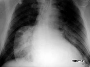
1.37 A metallic foil affixed as an electrode on the thorax produces a surprisingly distinct “shadow”.
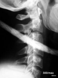
1.38 Isolated Cable Produces a Significant Artifact
38 XBBXHXINXKNXN
When the cable is at a large distance from the film, its depiction is greatly magnified and very blurred (geometric blurring).
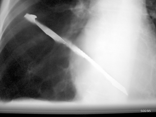
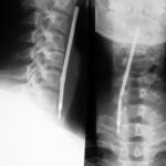
1.39 Appearance of a Ballpoint Pen in the Hypopharynx/Esophageal Inlet.
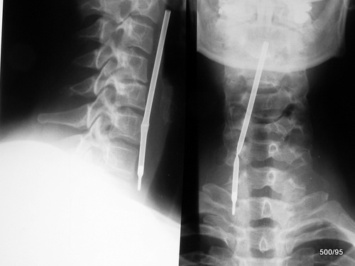
39 XBBXHXKJXQXPXW
The trachea is clear (see lateral image on the left). Uncomplicated extraction.
The following are some images of ingested foreign bodies.
On this topic, there is a worthwhile review article in the German Medical Journal (Deutsches Ärzteblatt) Volume 109 from 14.12.12 by Peter Ambe and colleagues from Düsseldorf. Since this article is sparsely illustrated, we can supplement it very well.
Ambe outlines the classification options for ingested foreign bodies.
- by size
- by surface (sharp vs. blunt)
- by material
- by properties (radiopaque +/-; chemically active +/-).
The most common ingested foreign bodies in adults are
- fish bones,
- followed by bones,
- followed by dental prostheses
Sung et al: Dig. Liver Disease 2011, 43; Chiu et al: A J Med Sci 2012, 343; Peng et al: Eur Arch Otorhinolaryngol 2012, 269.
1.39b. No foreign body. Density fracture at the base. Surgical repositioning and fixation with a long screw.
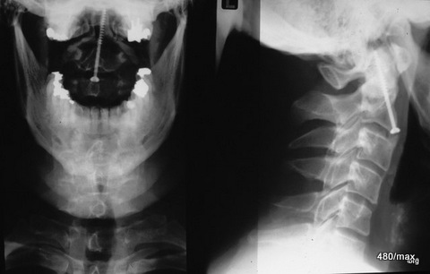
Now the atlas can no longer slide backwards and pinch the spinal cord.
1.40 Two (!) coins in the esophageal inlet.
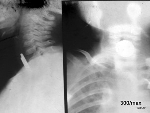
40 XBBXHXKJXQXPXW
A film from 1934 that is severely underexposed, with heavy scratches and other artifacts. The scratches can be light or, as shown here, black.
If the silver-containing film material is only lightly scraped off, a very fine light line is produced with a somewhat bolder black line parallel to it.
If the film material is completely scraped off, essentially wiped away, a distinct light scratch appears.
Apparently, very fine scratches tend to be black, while very broad, coarse scratches tend to be light.
The location of the coins is typical; if located in the glottis, foreign bodies of this shape would be arranged sagittally in the frontal image.
In the lateral view, they would be visible in the overview.
1.41 Does anything strike you about this thorax? Aspiration? Ingestion?
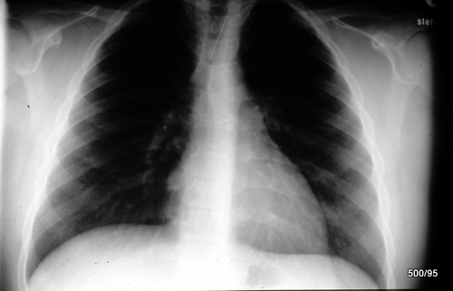
41 XBBXHXKJXQXPXW
Needle in the esophagus.
How can you rule out that the needle is in the trachea?
- Medical history
- Analysis of the image
- Second plane
- Breischluck?
I assume that we must first determine exactly what the patient reports and feels.
Then, a second plane appears to be a helpful document and should not delay timely extraction.
The image is inadequately displayed, no radiation protection.
1.42 82-year-old female patient; Confirmation of the suspicion of an ingested foreign body:
Es bestätigt sich ein abgebrochener Plastiklöffel, der sich noch im Magen befindet. Es wurde ein Breischluck gegeben. Man hätte das geschluckte Kontrastmittel stärker verdünnen können; . Ein Plastikbesteck stellt sich hell dar, wenn er von Magen- Luft umgeben ist; er stellt sich relativ dunkel dar, wenn es von einem dichten Kontrastmittel umgeben ist. Wahrscheinlich erscheint es auch in Wasser dunkel. Die orientierende Kontrastmittelgabe sollte mit einem wasserlöslichen Kontrastmittel erfolgen; sie soll eigentlich nur eine anatomische Orientierung bieten.
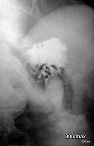
Hier sollte aus den deutschen Beitrag 1.44 BIld und Texxt rein!!
1.44a Detail Enlargement : Two Swallowed Bottle Caps.
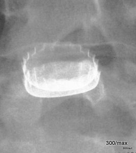
44 + 44a XBBXHXKJXT
During the follow-up examination one day later (right part of the image), the patient had swallowed a second bottle cap; it has conformed to the first one.
Enlargement of a section of the image from day 2. – Artifacts are sometimes also curiosities. (The latter would provide material for another topic).
1.45 Abdominal Overview in an Upright Position in a Child. Object? Location?
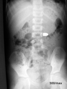
45 XBBXHXKJ. Ingested projectile (or is it a cartridge?) from a pistol. Likely still in the stomach. Incidentally: In the majority of cases of ingested foreign bodies, they pass naturally and without complications. In approximately 20% of cases, an endoscopy is necessary and advisable. In about 1% of cases, a surgical intervention is required.
In the present examination, gonadal shielding is missing; this causes issues with the Medical Authority.
1.46 Foreign Body in the Trachea?
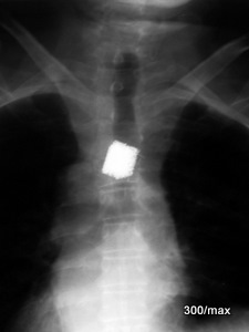
46 XBBXHXKJXLXPXW. Not in the trachea. The thorax x‑ray was not performed due to an emergency indication, but because of an unclear fever. The exposure is intentionally rendered dark here.
The patient can help. Condition following implantation of a titanium sponge in the area of a cleared bone metastasis of a thoracic vertebral body.
There is absolutely no clinical evidence of aspiration.
Purely morphologically: extension beyond the borders of the trachea. Some clips in the vicinity.
1.47 Follow-up many years after a vertebral fracture. What and where is the dense material?
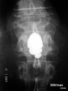
47 XBBXHXKJXPXW
Residuum from an examination performed many years ago. The indication was the question of a spinal canal stenosis and myelocompression due to a fracture of T12.
It involved an oily, hardly resorbable contrast medium: Duroliopaque. It provided an excellent contrast and moved freely in the cerebrospinal fluid space in accordance with its higher density.
It was virtually not resorbed, so that it had to be suctioned out after the examination, which was not always completely successful. The contrast medium moved freely in the CSF space. Over time, it frequently led to arachnoid irritation and was often trapped in basally adherent CSF spaces.
Today, we have better contrast agents and need them much less frequently, as there are also better methods that do not require contrast medium.
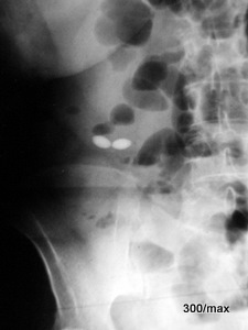
1.48 Where are the two pills located?
1.49 Where is the radiopaque, partially dissolved tablet material located?
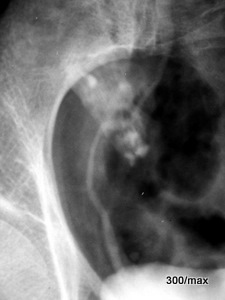
48 XBBXHXKNXS Upper Image
Is the subileus responsible for the fact that these tablets are still in the small intestine after 4 hours and have not yet dissolved?
Has any of our esteemed readers made an observation in this regard? See the article by Otte cited in the next case. There is no comprehensive
“list of tablet appearances in X-ray images.” This could be a great opportunity for diligent doctoral students—more than just a mini-atlas:
an analysis of the densities as a sum of individual components.
Would anyone dare to diagnose such an undissolved tablet using ultrasound?
The spatial assignment to the intestinal section should be relatively easy.
49 XBBXHXKN
The right ureter and bladder have been contrasted using excreted renal contrast medium. This incidentally illustrates
the spatial relationship between the ileocecal region and the right ureter.
Here, probably already in the distal ileum and cecum, there is still “partially dissolved” tablet material.
Related Research: In Deutsches Ärzteblatt, issue 110, volume 50 (2012), the previously cited excellent article on swallowed foreign bodies by Ambe P.
and colleagues was published.
This article was commented on by G. Bach. He explains that many, but not all tablets contain titanium dioxide as a “white pigment” or other additives with elements of a higher atomic number (bromine, iodine).
This explains their “density in the X-ray image” when they are not dissolved and therefore appear as solid structures.
He refers to his work: Bach AG…: 51-year-old patient found in a somnolent state. Dtsch med Wochenschr 2013; 138 S421.
Sonographic Detection
In the same discussion forum, A. Otte et al. advocate for the consistent use of ultrasound for the detection of foreign bodies.
They embedded various foreign bodies in gelatin and were able to detect them well using sonography.
Foreign Bodies in the Bladder
Several visitors have asked me whether I have an image of a foreign body in the bladder.
I do not currently have such an image available. Foreign bodies in the bladder can only be seen if they contain
calcium or metal. Otherwise, they would only be visible as filling defects in a contrast-filled bladder
(appearing as “black” on the X-ray).
1.50 Pelvic Overview. Examination Ordered by the Public Prosecutor. –
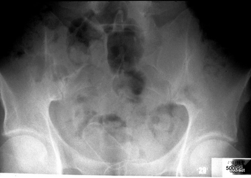
50 XBBXHXKJXLXUXPXW
In the small pelvis, there are at least two elongated structures with a distinctly bright rim and – compared to the soft tissues (i.e., relatively) – hypodense content: Packs with narcotics in the rectum.
Packs with narcotics in the rectum.
1.51 Example of unusual practices and patients who must seek our help due to the resulting hazards.
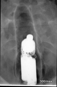
51 XBBXHXKJXQXPXW
Vibrating device in the rectum.
1.52 Fig.: Intravenous injection of Hg (mercury) with suicidal intent

1.52 XBBXHXKJXLXRXQXPXW.
Topogram of the thorax from a CT scan. Status post intravenous injection of mercury with suicidal intent.
The liquid metal has settled in droplet form in the capillaries of the lung and between the trabeculae of the right ventricle.
The patient survived the severe event. Even 4 years later, mercury was detectable in the pulmonary circulation. The systemic mercury poisoning was surprisingly mild.
In contrast to the dangerous inhalation of Hg vapors, the toxic effect of the large mercury droplets over the years was low, and the patient fared quite well.
With the kind permission of Prof. Eberhart Walter, Stuttgart/Tübingen
Chapter 2, Dark Artifacts
“I see something dark” is always a relative statement;
something appears dark due to the presence of neighboring brighter structures or when embedded in bright structures.
The classification into this Chapter 2 is often arbitrary. In several cases, it can be debated whether it is a light or dark artifact; whether it would belong in Chapter 1 or Chapter 2.
General rules:
Dark artifacts are usually caused by unwanted energy (light, X‑ray radiation, mechanical or thermal influence). Part of these dark artifacts therefore has nothing to do with X‑ray radiation.
2.01 Classical Light Incidence.
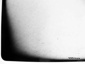
2.02 Bright or Dark Artifact?? Both Are Correct
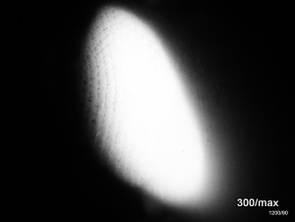
1XBBXDXELXFOXKNXO
The film was relatively protected in a drawer or box; the light incidence came from two edges.
The light incidence occurred before an X‑ray was taken. Of course, the film was developed, since it is the development that transforms the exposure into darkening.
Remarkably: In the area of the white field, where only very little light reached, there are fine black spots and dots. This is characteristic of the very low light incidence; it will concern us further in 03, 04, and 06. A covering with paper can play a role, but it does not have to.
2XBBXDXELXGXKNXNXO
With a large-area light incidence, a spot—a rounded area—remained shielded by an overlying fingertip; consequently, a white spot results.
It remains unclear to me the suggested fingerprint on the left edge of the white spot. Small amounts of a substance that a finger has imprinted may have initiated the development process and produced a dark structure??
Artifacts are very diverse. I have followed up on some observations; the resolution of some other problems I have left to my readers. Many mysteries of artifacts remain unsolved.
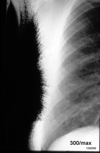
2.03 Light has apparently – very focused – hit an area on the left edge of the image.
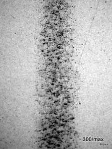
2.04 Discreet light incidence at fourfold magnification. Remarkable: coarse granularity.
3 XBBXDXELXFOXKN. Strangely, smaller amounts of the light energy radiated in a star-like pattern into the surrounding area. It remains a challenge to reconstruct such phenomena.
4 XBBXDXELXKNXL. This streak appears completely homogeneous when viewed from a distance of two meters; only upon closer inspection does one notice the mottled structure.
It remains to be determined whether a weak or tangential incidence of light (?) leads to such a relatively irregular film darkening. Did the light pass through paper? According to my reconstruction, that is unlikely.
It is still unclear!
2.05 Light incidence over several point-like defects in a shield and what else?
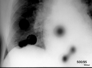
5 XBBXDXELXKN. It is likely that light from a not too small light source entered through small holes. Were there two light sources present, perhaps with different shapes?
In five of the black spots, one gets the impression that a core shadow and a penumbra are distinguishable.
Two of the spots are completely black and sharply delineated. Here, the intensity may have been so high that even the penumbra is deeply darkened. Or was this light source very small?
Thus, the size of the light source, its distance, the dose, and the size of the defects—all these factors may have played a role together.
2.06 A Provoked Artifact!
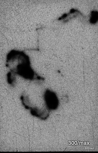
Trace of a firefly on a film!
The lively little insect did not fly into the darkroom by accident; it was invited to participate in this experiment. The slower its movement, the more intense the exposure. Individual short exposure streaks and dots are the expression of a very brief pause and flash. In addition, many dark scratches on the film and an unsettled texture are likely due to minimal diffuse lighting.
2.07 A Classic Artifact; it is difficult at first glance to determine whether it should be classified among the bright or dark art products.
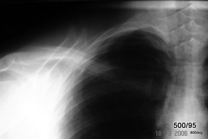
The artifact is not harmless, as it can lead to misdiagnoses: double exposure.
In any case, the film is noticeably dark. It is recognizable by the doubly depicted spinous processes, ribs, spinae scapulae (the spine of the scapula) and clavicles (collarbones).
The artifact can (as so often) be easily avoided.
2.08 Double Biliary Duct?
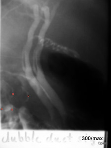
8 XBBXDXERXGXINXKN
“Dubble duct”. No; it is an artificial construct.
Double exposure. This results in a duplicated, laterally shifted depiction of the bile duct.
Bright or dark? This case could be attributed to either dark or bright artifacts.
A large number of the artifacts shown here are caused by organizational errors; they will be avoided in the future by changing the procedures.
2.09 An important case for everyone who no longer recognizes film-specific artifacts:

9 XBBXDXERXFDXKNXL
Aging of the unexposed film material.
Granular structure due to countless fine, spotty black artifacts; these probably arise from cosmic and terrestrial radiation and affect the entire film, as well as other films. Such film packs must be discarded.
If the baseline fog in the constancy test increases, one possible cause is this film aging.
2.10 Film Darkening Not Caused by Light??
Where Does the Mysterious Code Come From?
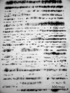
10 XBEXDXEEXKNXLXN. Probably due to an electrical discharge; here from the rollers of the developing machine. Not all of these cases are 100% confirmed!
The classic example of such electrical discharges are the black “trees”, as shown in the article “15 Colorful X‑ray Images”.
Also, the characteristic discharge emanating from a fingertip is demonstrated there.
The manifestation shown here is a rarity. Many preconditions have to come together; among these, high air dryness is decisive.
A wet towel on the heater or another method to humidify the air can help.
How can one prove that these are not light but electrical artifacts? Scrape off a small piece of emulsion from both sides of the double-coated film: Light affects both coatings (XBB), electrical energy only one side (XBE).
We are glad that with digital techniques we no longer have to deal with such film-specific artifacts.
2.11 Even electrical discharges come in an enormous variety of forms.
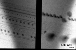
11 XBEXDXEEXKNXS
A general rule applies: If an artifact exhibits only a slight manifestation, its classification as an artifact becomes more difficult, and thereby the risk of misinterpretation increases.
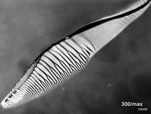
2.13 Parasite?
2.14 What happened here?
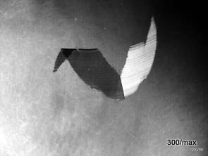
13 XBEXDXEHXGXKNXLXN.
In this case, the diagnosis is easier to establish by palpation than by inspection: melting of the film emulsion in a defective drying unit. It was observed during a humanitarian mission. A rare and therefore valuable artifact for collectors. Rarity does not mean that the phenomenon cannot occur again tomorrow. If it does, we will immediately classify it. This image was colorized in Case 15.
There are analogous cases in the (not entirely serious) literature that deliberately treat artifacts with humor: Hoeppli, R.: Parasites and parasitic infections in early medicine. Singapore: University of Malaya, 1959; 164.
Wendth, AJ, Sarah Reed-Esper: Inner Visions. Radiographics 13, 1264.
14 XBEXDXEMXGXKN. Emulsion scraped off on one side, displaced, flipped, and then re-applied to the film.
Compared to thermal damage (13), such mechanical noxae are not uncommon.
The film is double-coated (with few exceptions). This is done to save dose. If there is a defect on one side of the coating, the impression of a bright area is created, while another exposed coating remains intact. As always, the impression is relative.
In the case of scratches, the film emulsion is often not removed but only displaced; at the site of displacement, three coatings accumulate. In addition to this broad lifting of the emulsion, a field with the impression of strong darkening arises (on the left side).Thus, such artifacts consist of both a bright and a dark component. This will also be demonstrated in another form in the next case:
2.15 – Very old thorax X‑ray. Scratches on the film. Are they bright or dark?
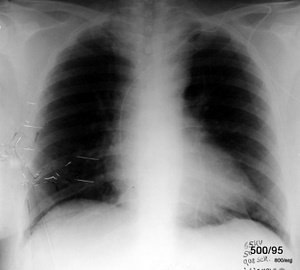
15 XBEXDXEMXGXKNXO. The notes in the lower right corner (overlapped by my own notes) reveal how poorly the settings were chosen back then. The use of 65 kV is nowadays prohibited for radiation protection reasons (see the “Radiation Protection” article). Typical for low kV settings are high mAs values. Progress is often a real gain.
It is also progress that we are no longer dependent on chemical processing.
The records reveal: the catheter is compressed and was corrected by retraction. Rightly, you may ask: Why are the outer subfields so unevenly dense? Is there a pathological opacity in the left subfield? I do not believe so. It is probably due to asymmetrical soft tissues, both the breast shadow and the thoracic wall.
This film was treated carelessly and, especially in the lower left quadrant, creased and scratched. By now, you know that scratches can be partially black and partially light. These are examples of extreme artifacts, resulting from the imprudent handling of documents.
2.16 What happened here?
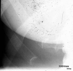
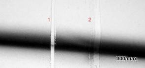
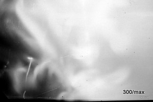
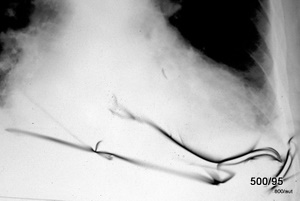
20 XBEXDXEMXFOXKN
Initially, an unusual light incidence was assumed; later (in light of the experiments 2.7 b + c, 2.18 and 19) this was corrected to: “Mechanical alteration prior to development“. One can still see and feel the deformation on the film. Not the entire pack, but the individual film was unusable.
It is undisputed that many artifacts can lead to misdiagnoses (small fracture lines, etc.) especially when they are only subtly manifested and their nature is not immediately recognizable.
2.21 Lichteinfall oder mechanische Alteration??
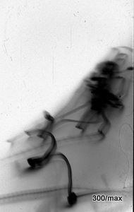
21 XBBXDXELXFOXKN
Despite the similarity to the previous case, we suspect here a focused light source that moved (relatively) with respect to the film. The safety is ensured by the “scratch test” (see 19). This is analogous to the case of the firefly, where an exposure was deliberately produced. Mechanical damage to the film as the primary diagnosis is excluded (although smaller scratches are also present).
2.22 X‑ray from the year 1932. A case for professionals!
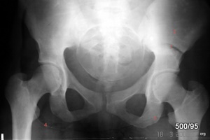
22 XBXDXEXKXPXW
We know nothing about the patient, the indication, or the imaging parameters.
One should be very cautious when evaluating such images that are completely
disassociated from their clinical (and technical) context. The quality is poor;
this was not uncommon in these early days.
At first glance, the head of a child appears completely uncontroversial. The finding jumps out.
Upon closer inspection, however, there are so many artifacts that even the “pregnancy” becomes very uncertain.
Unfortunately, the red numbering of my problems is blurred.
Deviously, artifact 1 — the left pelvic half 4 cm above the acetabular dome — is so precisely confined to the bone and suggests a remodeling zone. (See Article 01.)
The localization and the lack of an osteoplastic reaction do not at all fit this diagnosis.
“4” is a downward-convex line between the right lesser trochanter and the right ischial tuberosity: certainly also artificial.
Overall, the bony structure is very poorly depicted; or is there truly an underlying disease?
The left hip is rotated outward very carelessly. The left ischium, near the hip joint, appears somewhat raised,
perhaps even the upper pubic ramus near the hip joint? Could it be a residue of a fracture or a remodeling zone?
We venture the hypothesis: Very poor exposure full of artifacts.
I, WGHS, work on radiology in the first half of the 20th century; it was actually at a high technical level.
Only a few important aspects of radiation protection were insufficiently considered at that time.
It is unusual for images from that era to exhibit so many artifacts.
What will people say in a few decades about our workplace and our time?
2.23 From the Era of Chemical Film Processing. The bright image areas are particularly obscured and thereby darkened. One must know this!
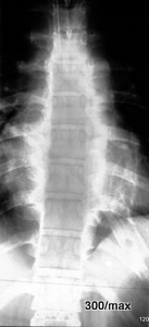
23 XBXDXEXKXPXW
Undoubtedly a fixation error (namely, insufficient fixation).
In the exposed areas of the image, the chemical process of film darkening has occurred, which becomes visible during development. Silver is released, and the film turns black!
What happens in the less or unexposed areas? Here, the underexposed silver salt must be precipitated so that the film becomes nicely bright and transparent. This also ensures that the film becomes stable and that the salt, which can still react chemically, is no longer present in the film. This is an important function of fixation. In this case, the fixation did not work; this unexposed silver salt was not precipitated and, through a paradoxical chemical reaction, has darkened the particularly minimally exposed parts of the film.
The most striking aspect of this phenomenon is the reversal of the light-dark scheme. The normally brighter base plates and arcuate roots are darker than the surrounding areas.
Dark structures, such as the lung, remain unremarkable. The developer was intact—it darkens the exposed structures. Only the unexposed silver salt was not washed out due to insufficient fixation.
In short: This image example represents a faulty development process with an intact developer but insufficient fixation.
An important case. One must know this artifact in order to react in time and correctly.
Review: What does insufficient development look like? Primarily, the dark areas of the film would not be dark enough. The disturbance would not affect the bright areas but rather the dark areas of the film.
In the constancy test, an elevation of the base fog is observed in the first case discussed; the described phenomenon is one of the possible causes.
2.24 X‑ray Film Processing. This is what an unexposed film looks like??
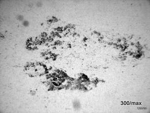
24 XBEXDXFTXKN. In the water bath, algae grew. These were pressed onto the film during washing and drying. In effect, a herbarium was created. A disturbing, expensive artifact but – as so often – one that is easy to correct.
The more specific our orders to the technical staff, the more time can be saved.
2.25 Die Entwicklermaschiene fördert diesen Film zutage. Ursache?
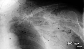
– A triviality. Who would willingly display such a disastrously failed product? Not for radiological diagnosis, but rather as a mud-slinging.
2.26 Black “Hole in the Heart”?
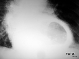
26 XBEXDXPXKJXLXPXW
This is a paraesophageal, air-filled hernia behind the heart.
It should rather be classified under the “Pathology” article. If I were to classify this case among the artifacts, then it could also have found its place among the “bright” ones.
What appears dark to us is normal exposure. Under normal circumstances, lung tissue would predominate. The wall layers of the hernia have displaced lung and produce a thick white ring = an opacity in the strict radiological sense; in contrast to this white ring, the relatively dark center appears to us like a “black hole in the heart.” As is often the case, the impression in the X‑ray image is relative.
2.27 Dark Structure Projected onto the Small Pelvis?
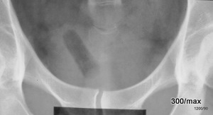
27 XBBXDXPXLXOXW
The image is only worth showing because it educates our theoretical understanding: it clearly demonstrates how arbitrary the classification into black or white artifacts can be. Tampon in the X‑ray image.
Like a red thread running through the entire presentation, the rule is: The same object is depicted very differently depending on whether it is surrounded by air or by soft tissue.
An example can be found in the article “Radiation Protection 09”; a part of a light bulb in the intestine appears predominantly black. –
The tissue shown here is still very air-filled (it consists of much air and little tissue). Here, it appears black; it displaces tissue, removing it from radiation attenuation; it removes more than it contributes. If this structure were superimposed on the soft tissues externally, a (small) increase in density would result; it would appear as a (more or less) brighter spot.
For this issue, see also the following observation of a traumatically incorporated foreign body, 28)
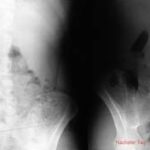
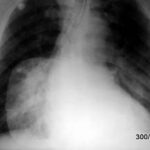
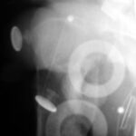
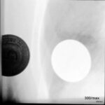










Comment on 1.02 XBBXHXFLXINXKNXL
Upon closer inspection, the strip is light but not as maximally white as a completely unexposed film. In this strip, the film blackening is largely (though not entirely) removed.
In the immediate vicinity of the strip, there is a zone de Unschärfe of the thoracic structures.
Explanation: In the old conventional process, films are predominantly exposed by the intensifying screens. This foreign body at least removes the light emanating from one of the two screens; that half of the light is not available for film blackening.
Moreover, it lifts the screen away from the film in its vicinity. As a result, a blurring occurs. More precisely: One of the screens produces a sharp image, the other a blurry image; the sum is a deterioration of the usual image sharpness.
HG Schmitt also detected hairs on the film. Therefore, he asked all the staff for a hair and tested whether a hair, based on its thickness, could be attributed to a specific person.